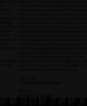
Labels:text | screenshot | font | black | black and white | document OCR: enal gland : Fig. 23.12 Conn's adenoma in the left ad hal demonstrated by venography (with non ws the venogram for comparison). Film (a) sho h's typical venographic appearance of a Con drenal veinadenoma. The catheter tip is in the left a etched and contrast medium has filled veins st ole of the around a 1 cm tumour in the superior p mation adrenal gland. Blood for aldosterone est rior to sation of should be taken from the adrenal vein venography because of the risk of extrav Even contrast medium during that procedure adrenal careful venography carries a small risk c infarction which could result in adrenal insufficiency if both adrenal glands are e is rarely compromised. Adrenal venography alo theter carried out today, except to identify the c position during venous sampling. See earlier: ...